Tingling in the hands during training, a “dead foot” mid-run, or unexplained weakness in grip or push-off strength is often dismissed as a minor issue. In sport, these symptoms can reflect peripheral nerve irritation or entrapment, where mechanical compression or repetitive load affects neural function.
Unlike muscle injuries, nerve-related conditions often develop gradually and present with a mix of sensory symptoms (tingling, burning, numbness) and motor changes (weakness, altered coordination). Because of this, they are frequently misinterpreted or overlooked in early stages.
Most cases are managed conservatively. Physiotherapy plays a central role in reducing irritability, restoring movement tolerance, and guiding a structured return to sport through progressive loading.
Some of the common peripheral nerve conditions seen in athletes include:
Carpal Tunnel Syndrome
Cubital Tunnel Syndrome
Radial Nerve Palsy or Irritation
Common Peroneal Nerve Irritation
Tarsal Tunnel Syndrome
Obturator Nerve Entrapment
Meralgia Paresthetica
Carpal Tunnel Syndrome (Median Nerve – Wrist)
Median nerve compression at the wrist is common in cycling, climbing, gymnastics, and resistance training.
Presentation
Tingling in thumb, index, and middle fingers
Night symptoms or waking with hand discomfort
Reduced grip endurance
Hand fatigue with repetitive loading
Physiotherapy approach
Management focuses on reducing local irritation while maintaining upper limb capacity:
Load modification (grip volume and wrist positioning)
Median nerve mobility work
Forearm soft tissue and mobility techniques
Progressive wrist and hand strengthening
Shoulder and scapular control to reduce distal load
Cubital Tunnel Syndrome (Ulnar Nerve – Elbow)
Ulnar nerve irritation at the elbow is common in throwing athletes, cyclists, and gym-based training involving sustained elbow flexion.
Presentation
Tingling in ring and little fingers
Symptoms aggravated by prolonged elbow bending
Medial elbow ache
Reduced hand dexterity or coordination
Physiotherapy approach
Reduce sustained elbow flexion positions (especially overnight)
Taping or positioning strategies if required
Ulnar nerve gliding exercises
Forearm strengthening with graded exposure
Scapular and postural control work
Radial Nerve Palsy or Irritation (Upper Arm)
Radial nerve dysfunction can occur through compression or traction in contact sports, falls, or heavy resistance training.
Presentation
Weak wrist and finger extension
Difficulty releasing grip
Numbness over the dorsum of the hand
Physiotherapy approach
Rehab is guided by severity and irritability:
Protection or splinting if required
Gradual activation of wrist and finger extensors
Neural mobility exercises when tolerated
Upper limb kinetic chain strengthening
Functional reloading into gripping tasks
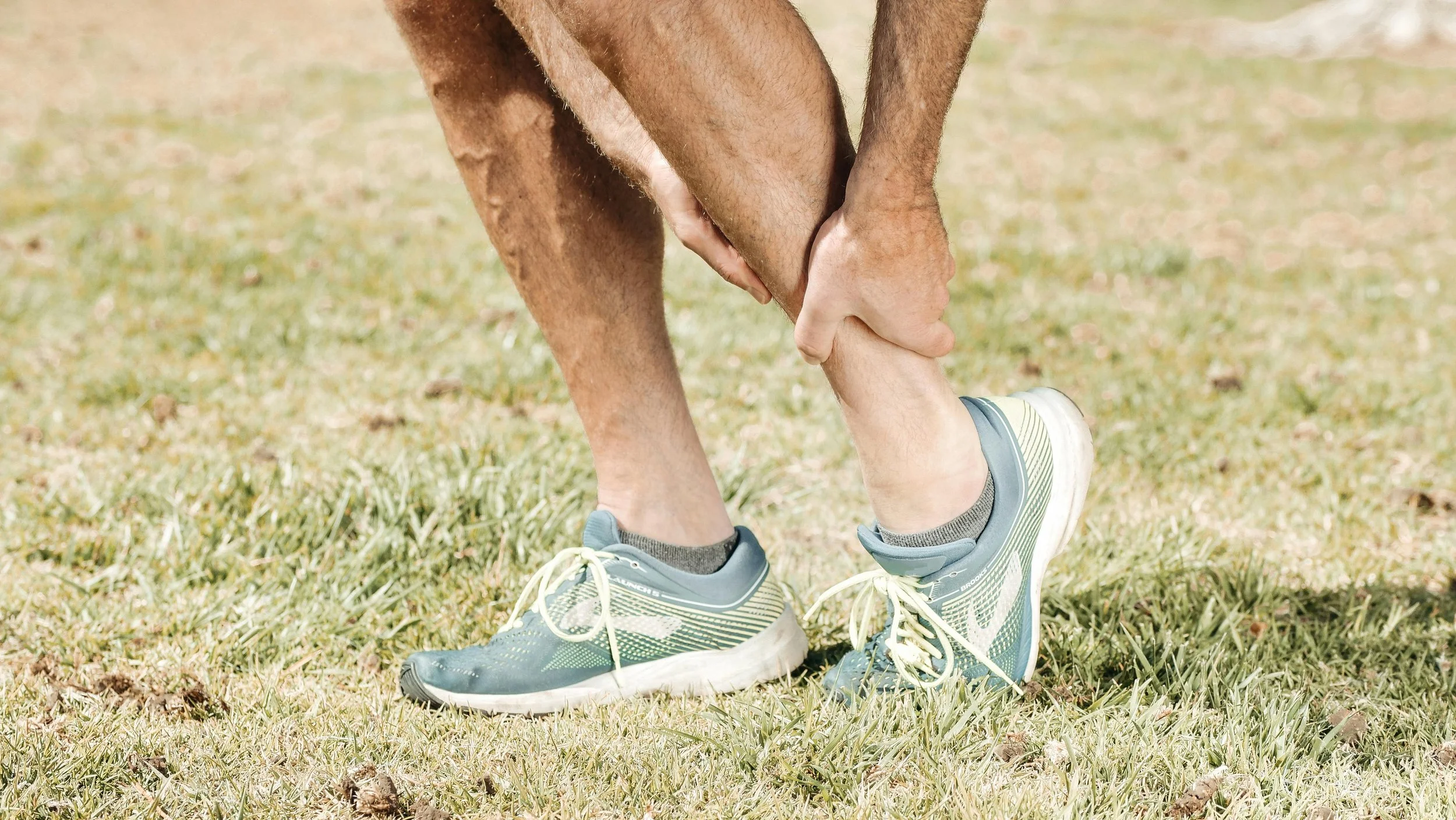
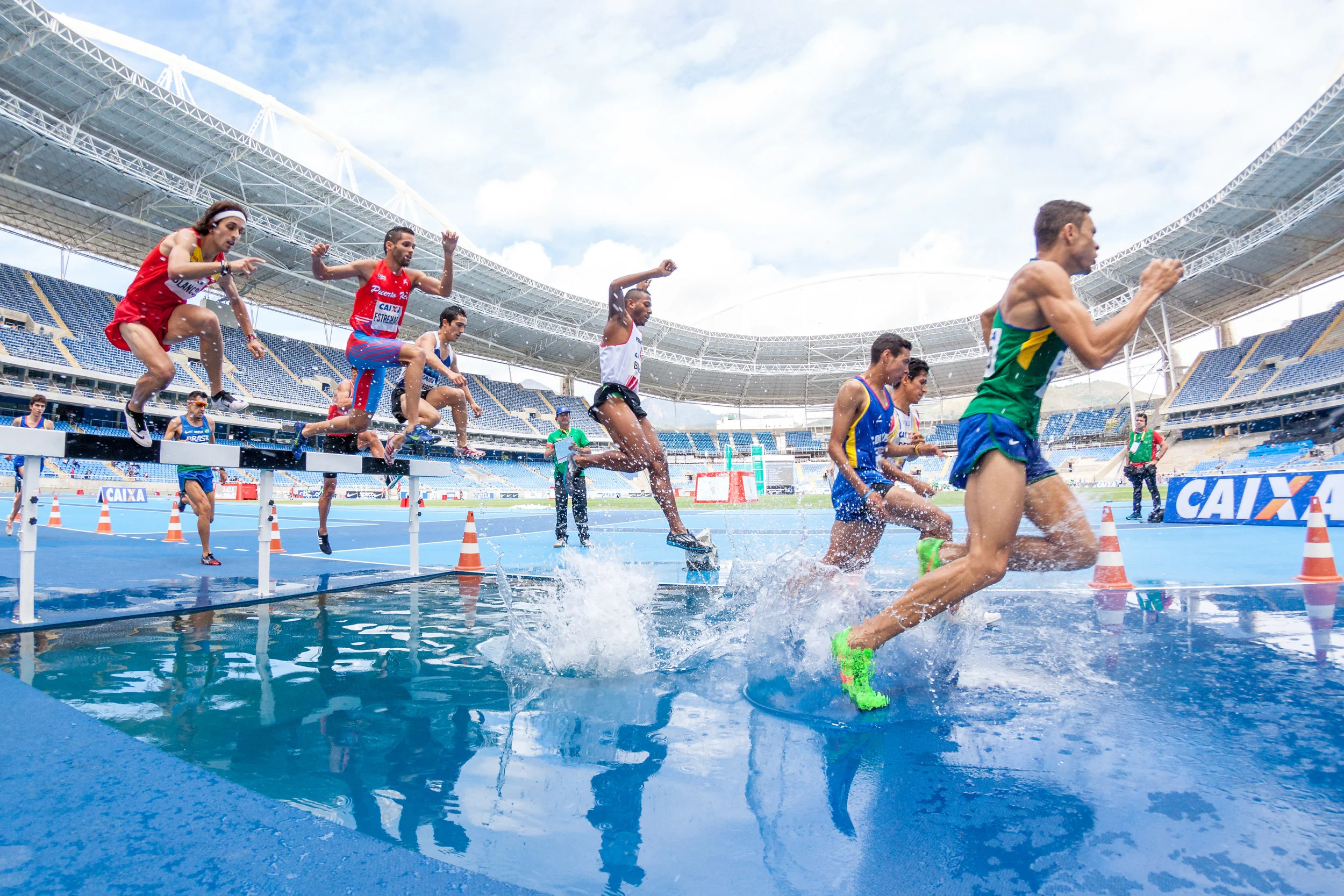
Common Peroneal Nerve Irritation (Fibular Head – Knee)
The common peroneal nerve is vulnerable as it wraps around the fibular head, particularly in running and field sports.
Presentation
Foot drop or toe drag
Dorsal foot or lateral shin numbness
Weak dorsiflexion
Altered running gait mechanics
Physiotherapy approach
Identify and remove external compression sources
Bracing or AFO fitting
Gait retraining and running re-education
Strengthening of dorsiflexors and evertors
Balance and proprioceptive training
Hip and knee control to reduce distal overload
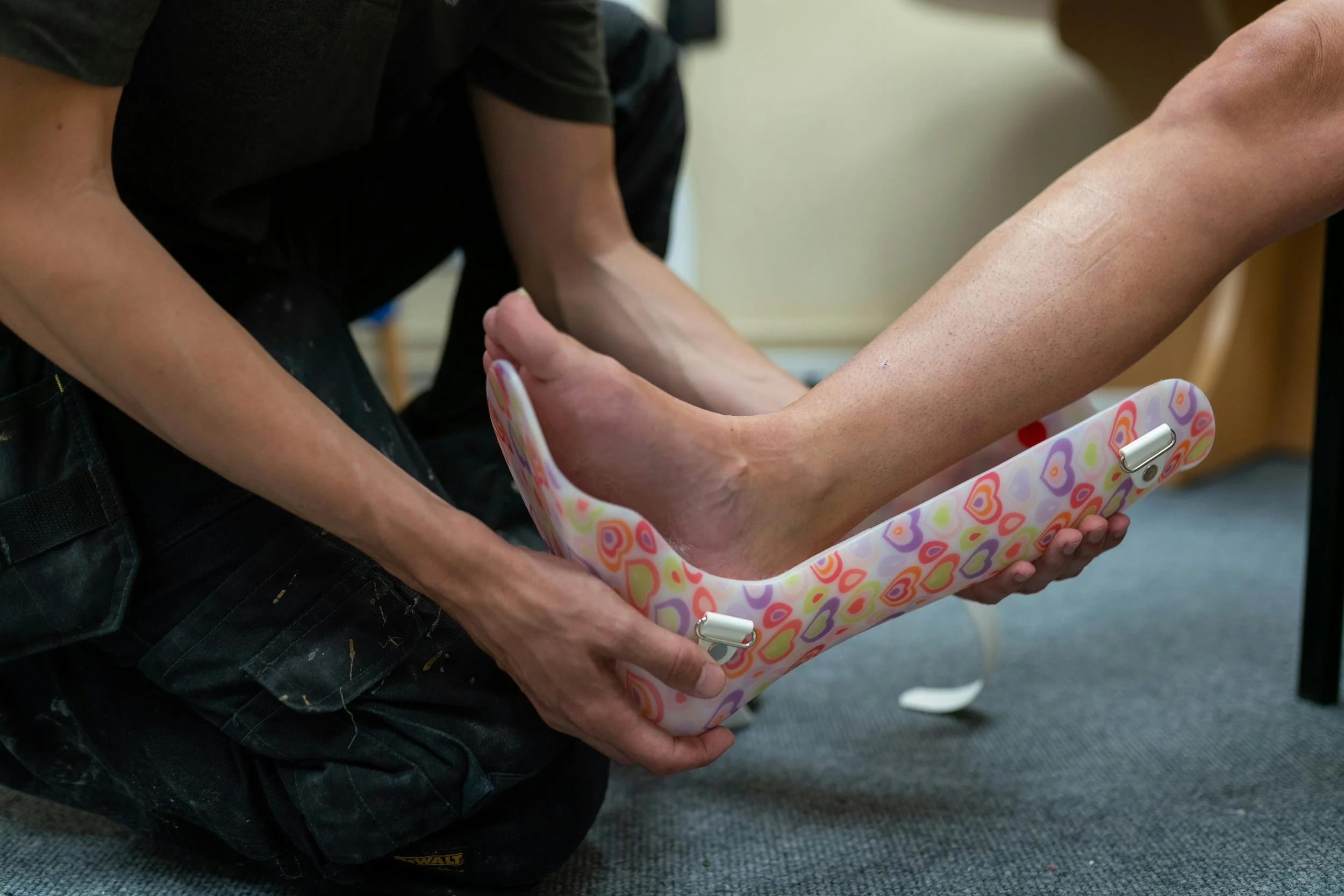
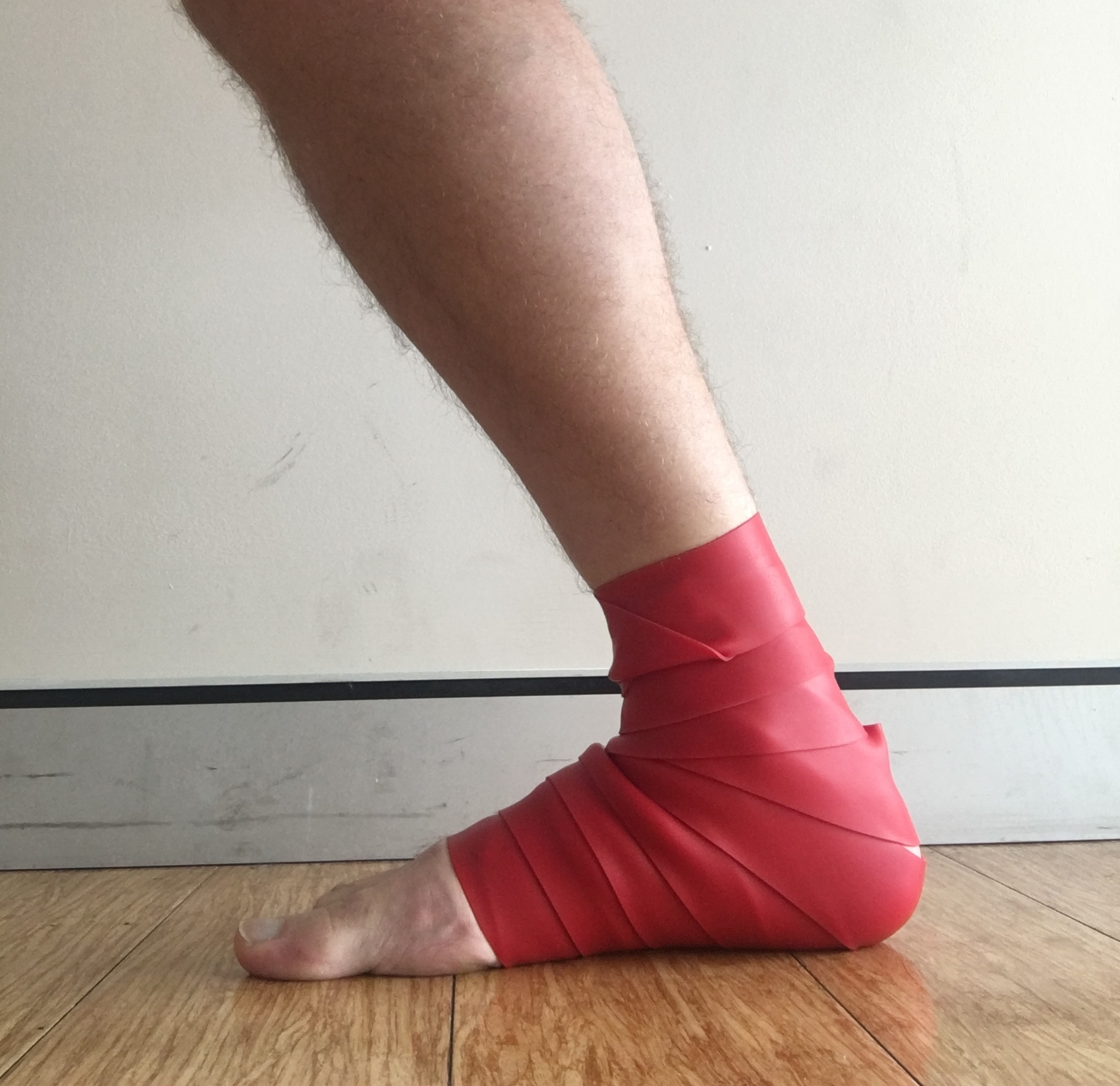
Tarsal Tunnel Syndrome (Tibial Nerve – Ankle)
Tibial nerve compression behind the medial malleolus is commonly seen in runners and jumping athletes.
Presentation
Burning or tingling in the sole of the foot
Medial ankle discomfort
Symptoms worse with running or prolonged standing
Often mistaken for plantar fascia-related pain
Physiotherapy approach
Load modification (running volume and intensity)
Calf and posterior chain strength and mobility work
Intrinsic foot muscle strengthening
Gait and biomechanical retraining
Tibial nerve mobility techniques (selected cases)
Obturator Nerve Entrapment (Groin / Inner Thigh)
Less common but important in multidirectional and kicking sports.
Presentation
Deep medial thigh ache
Pain with resisted adduction
Reduced adductor strength and endurance
Symptoms during cutting or sprinting
Physiotherapy approach
Reduce adductor load in early stages
Progressive adductor strengthening (isometric → isotonic → eccentric)
Hip and pelvic stability training
Soft tissue techniques to adductors
Movement retraining for change of direction
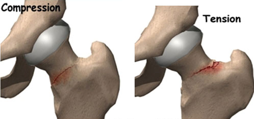
Lateral Femoral Cutaneous Nerve Entrapment (Meralgia Paresthetica)
Compression of the LFCN under the inguinal ligament, often related to repetitive hip flexion or external compression from clothes or equipment.
Presentation
Burning or tingling over outer thigh
Purely sensory symptoms (no weakness)
Worse with running, cycling, or tight clothing
Relief with standing or hip extension
Physiotherapy approach
Remove external compression (clothing, belts, equipment)
Reduce repetitive hip flexion load
Hip flexor mobility work
Lumbopelvic control training
Gradual desensitisation and return to activity
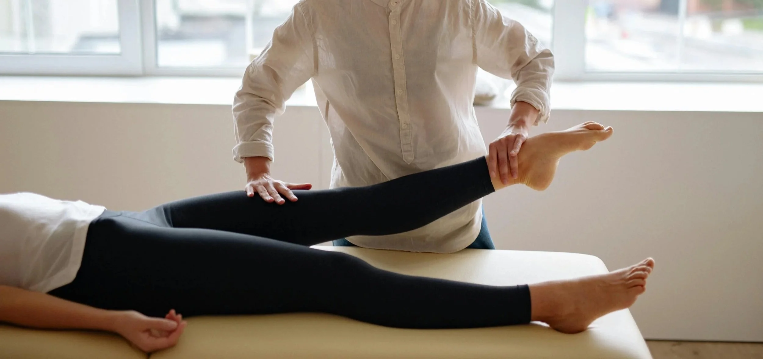
The Role of Physiotherapy
Peripheral nerve injuries are rarely isolated problems. They reflect a combination of load sensitivity, movement patterns, and mechanical irritation.
Physiotherapy management focuses on:
Load modification without complete rest
Improving neural mobility and tolerance
Restoring strength across the kinetic chain
Addressing contributing movement dysfunction
Progressive return to sport based on symptom response
Nerve tissue is adaptable but sensitive to both overload and under-loading. Successful rehabilitation requires a balance between reducing irritability and maintaining movement capacity.
Summary
Peripheral nerve injuries such as carpal tunnel syndrome, cubital tunnel syndrome, radial nerve irritation, common peroneal nerve dysfunction, tarsal tunnel syndrome, obturator nerve entrapment, and meralgia paresthetica are all relevant in athletic populations.
They often present subtly, but can significantly affect performance if not recognised early.
With appropriate physiotherapy management, most athletes respond well to conservative care and return to full sport through structured, progressive loading and movement retraining.
Further reading/references
Keith, M. W., Masear, V., Chung, K. C., Maupin, K., Andary, M., Amadio, P. C., … Yao, J. (2009). AAOS clinical practice guideline on diagnosis of carpal tunnel syndrome. Journal of the American Academy of Orthopaedic Surgeons, 17(6), 397–405. https://doi.org/10.5435/00124635-200906000-00007
Nakashian, M. N., Ireland, D. C., & Kane, P. M. (2020). Cubital tunnel syndrome: Current concepts. Orthopedic Clinics of North America, 51(3), 475–486. https://doi.org/10.1016/j.ocl.2020.03.009
Hölmich, P., Uhrskou, P., Ulnits, L., Kanstrup, I. L., Nielsen, M. B., Bjerg, A. M., Krogsgaard, K., & Jensen, J. (2010). Effectiveness of active physical training as treatment for long-standing adductor-related groin pain in athletes: Randomised trial. The Lancet, 353(9151), 439–443. https://doi.org/10.1016/S0140-6736(98)03340-6
Bramah, C., Preece, S. J., Gill, N., & Herrington, L. (2018). Is there a pathological relationship in running-related injuries? A systematic review of biomechanics and load management. Sports Medicine, 48(12), 2781–2798. https://doi.org/10.1177/0363546518793657
Staff, P. (2018). Meralgia paresthetica: Clinical features and management. StatPearls Publishing. https://www.ncbi.nlm.nih.gov/books/NBK557735/
Campbell, W. W. (2008). Evaluation and management of peripheral nerve injury. Clinical Neurophysiology, 119(9), 1951–1965. https://doi.org/10.1016/j.clinph.2008.03.018